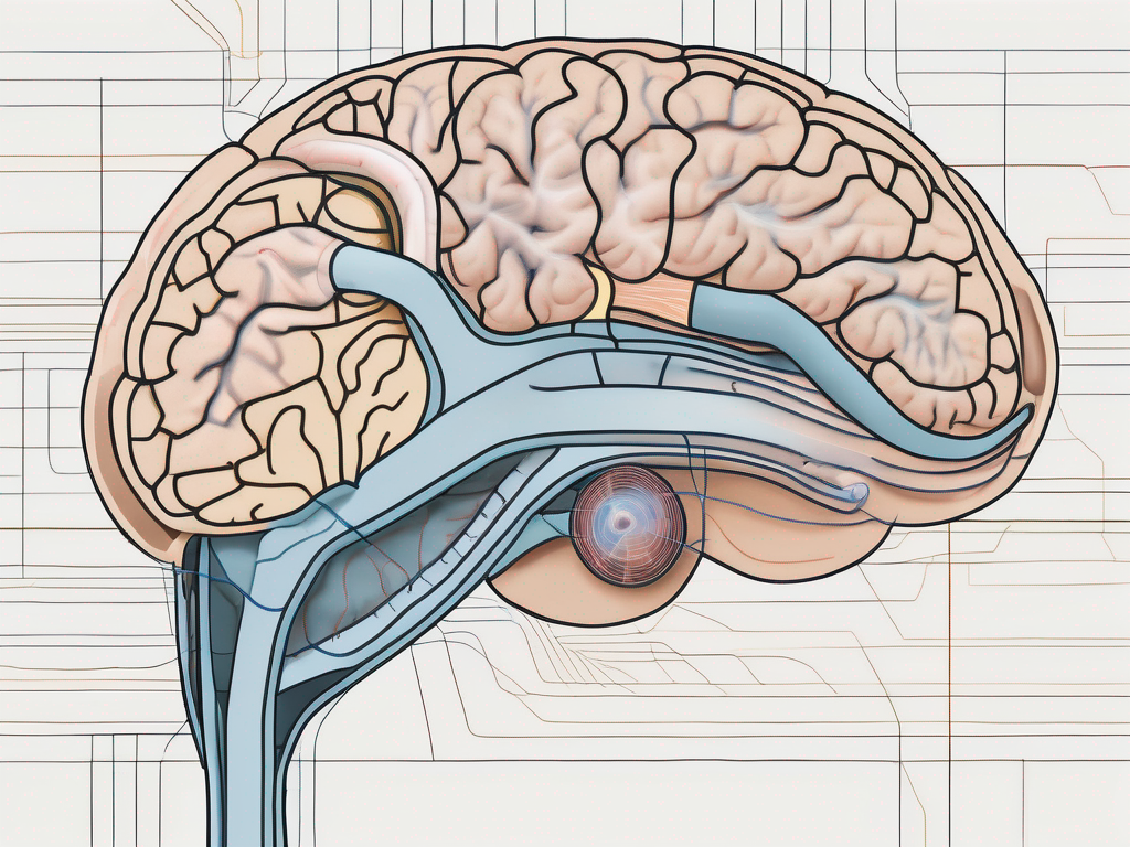
The vestibulocochlear nerve, also known as the eighth cranial nerve, plays a crucial role in our auditory and vestibular systems. It is responsible for transmitting sensory information related to hearing and balance from the ear to the brain. In order to fully understand the function and anatomy of this important nerve, it is essential to explore where its cell bodies are located.
Understanding the Vestibulocochlear Nerve
The vestibulocochlear nerve is a complex structure that consists of two main branches – the vestibular branch and the cochlear branch. The vestibular branch carries information related to balance and spatial orientation, whereas the cochlear branch is responsible for transmitting auditory signals.
The vestibulocochlear nerve, also known as the eighth cranial nerve, is a crucial component of our sensory system. It allows us to perceive and interpret sound, as well as maintain our balance and spatial awareness. Let’s delve deeper into the anatomy and function of this fascinating nerve.
Anatomy of the Vestibulocochlear Nerve
The cell bodies of the vestibulocochlear nerve are located in specialized clusters within the brainstem. Specifically, the vestibular branch originates from the vestibular ganglion, a group of cell bodies located within the internal auditory canal. This ganglion serves as the starting point for the transmission of balance-related information.
On the other hand, the cochlear branch arises from the spiral ganglion, which is situated within the cochlea of the inner ear. This ganglion is responsible for transmitting auditory signals to the brainstem, where they are further processed and interpreted as sound.
It is important to note that the vestibulocochlear nerve is a paired structure, meaning that there is one nerve on each side of the head. The cell bodies of the vestibular branch and the cochlear branch can be found on both the left and right sides of the brainstem. This bilateral arrangement ensures that our auditory and balance systems work in harmony to provide us with a comprehensive sensory experience.
Function of the Vestibulocochlear Nerve
The vestibulocochlear nerve plays a vital role in our ability to perceive and interpret sound and maintain balance. When sound waves enter the ear, they are converted into electrical signals by the sensory cells within the cochlea. These electrical signals are then carried by the cochlear branch of the vestibulocochlear nerve to the brainstem, where they are further processed and interpreted as sound.
Similarly, when we move or change positions, the vestibular branch of the vestibulocochlear nerve senses the movement and transmits this information to the brain for processing. This allows us to maintain our balance and adjust our body position accordingly. The vestibular branch is responsible for detecting changes in head position, rotational movements, and linear acceleration, ensuring that we can navigate our environment safely and efficiently.
Moreover, the vestibulocochlear nerve is intricately connected to other structures within the central nervous system. It receives inputs from various sensory systems, such as the visual and proprioceptive systems, to create a comprehensive picture of our surroundings. This integration of sensory information enables us to perceive the world around us accurately and make precise movements.
In conclusion, the vestibulocochlear nerve is a remarkable structure that allows us to experience the richness of sound and maintain our balance. Its complex anatomy and intricate connections within the central nervous system highlight the sophistication of our sensory systems. Understanding the vestibulocochlear nerve provides us with insights into the intricacies of human perception and the remarkable capabilities of our brain.
Location of Cell Bodies in the Vestibulocochlear Nerve
The vestibulocochlear nerve, also known as the eighth cranial nerve, is a crucial component of our auditory and vestibular systems. It is responsible for transmitting sensory information related to hearing and balance from the inner ear to the brainstem. The cell bodies of this nerve are located within specific structures in the brainstem, namely the vestibular ganglion and the spiral ganglion.
The vestibular ganglion, located within the inner ear, contains the cell bodies of the vestibular branch of the vestibulocochlear nerve. These cell bodies are responsible for processing sensory information related to balance and spatial orientation. They receive signals from the vestibular hair cells, specialized sensory cells within the inner ear that detect changes in head position and movement.
The spiral ganglion, on the other hand, is situated within the cochlea, the snail-shaped structure of the inner ear responsible for hearing. This ganglion contains the cell bodies of the cochlear branch of the vestibulocochlear nerve. These cell bodies receive and process auditory signals from the hair cells of the cochlea, which convert sound vibrations into electrical signals.
The Role of Cell Bodies in Nerve Function
The cell bodies within the vestibular ganglion and the spiral ganglion play a vital role in the proper functioning of the vestibulocochlear nerve. They act as the initial processing centers for sensory information received from the ear. These cell bodies receive electrical signals from the hair cells and convert them into a language that the brain can understand.
Once the sensory information is processed within these cell bodies, it is transmitted as electrical signals along the nerve fibers of the vestibulocochlear nerve. These nerve fibers extend from the ganglia and travel through the internal auditory canal, eventually reaching the brainstem.
Within the brainstem, the electrical signals are further processed and interpreted by various structures, including the cochlear nuclei and the vestibular nuclei. These structures analyze the signals and contribute to our perception of sound and maintenance of balance.
Additionally, the cell bodies within the vestibular ganglion and the spiral ganglion are responsible for the maintenance and regulation of the nerve fibers that make up the vestibulocochlear nerve. They ensure that the nerve is able to transmit information efficiently and accurately, allowing us to hear and maintain balance effectively.
How Cell Bodies Contribute to Hearing and Balance
The cell bodies within the vestibulocochlear nerve play a crucial role in our ability to hear and maintain balance. Through their intricate connections and communication with other structures within the ear and the brainstem, these cell bodies ensure that sensory information related to hearing and balance is accurately transmitted to the brain for interpretation.
When sound waves enter the ear, they cause the hair cells in the cochlea to vibrate. These vibrations are converted into electrical signals by the hair cells, which are then transmitted to the cell bodies within the spiral ganglion. These cell bodies process the signals and relay them to the brainstem, where they are further analyzed and interpreted as sound.
Similarly, when we move or change our head position, the vestibular hair cells within the inner ear detect these changes and send signals to the cell bodies within the vestibular ganglion. These cell bodies process the signals and transmit them to the brainstem, allowing us to maintain balance and spatial orientation.
Damage or dysfunction of the cell bodies within the vestibulocochlear nerve can lead to various hearing and balance disorders. Conditions such as hearing loss, dizziness, and vertigo can all result from problems in the processing and transmission of sensory information by these cell bodies. Therefore, the health and proper functioning of these cell bodies are crucial for our overall auditory and vestibular well-being.
Disorders Related to the Vestibulocochlear Nerve
Disorders affecting the vestibulocochlear nerve can have a significant impact on an individual’s quality of life. The vestibulocochlear nerve, also known as the eighth cranial nerve, is responsible for transmitting sensory information from the inner ear to the brain. It plays a crucial role in our ability to hear and maintain balance.
When this nerve is affected by a disorder, it can lead to a range of symptoms that can greatly disrupt daily life. It is essential to recognize the symptoms associated with these disorders and understand the available treatment options to provide relief and improve overall well-being.
Symptoms of Vestibulocochlear Nerve Disorders
Vestibulocochlear nerve disorders can manifest in various ways, depending on the specific condition and the extent of the damage. Common symptoms include:
- Hearing loss: Individuals may experience partial or complete hearing loss in one or both ears.
- Tinnitus: A persistent ringing, buzzing, or hissing sound in the ears.
- Vertigo: A sensation of spinning or dizziness, often accompanied by nausea.
- Dizziness: A feeling of lightheadedness or unsteadiness.
- Problems with balance: Difficulties in maintaining stability and coordination.
If you experience any of these symptoms, it is important to consult with a healthcare professional for a proper diagnosis. They will conduct a thorough examination and may recommend additional tests, such as audiometry or imaging studies, to determine the underlying cause of your symptoms.
Diagnosing the specific vestibulocochlear nerve disorder is crucial for developing an appropriate treatment plan and managing the condition effectively.
Treatment Options for Vestibulocochlear Nerve Disorders
The treatment of vestibulocochlear nerve disorders depends on the specific condition and its severity. In some cases, the underlying cause can be addressed, leading to an improvement or resolution of symptoms.
For example, if hearing loss is due to a blockage in the ear canal, removing the blockage may restore hearing. Similarly, if the disorder is caused by an infection, appropriate antibiotics can help clear the infection and alleviate symptoms.
In other cases, managing the symptoms and promoting rehabilitation may be the main focus of treatment. This can include strategies such as:
- Physical therapy: Specialized exercises and techniques to improve balance and coordination.
- Vestibular rehabilitation exercises: Targeted exercises to retrain the brain to interpret and respond to vestibular signals.
- Hearing aids: Devices that amplify sound and improve hearing for individuals with hearing loss.
- Cochlear implants: Surgically implanted devices that bypass damaged parts of the inner ear and directly stimulate the auditory nerve, providing a sense of sound.
It is important to note that treatment options for vestibulocochlear nerve disorders should be determined by healthcare professionals who specialize in this field. They have the expertise and knowledge to provide individualized recommendations based on your specific condition and needs.
Furthermore, ongoing research and advancements in medical technology continue to expand treatment options for these disorders. It is essential to stay informed and consult with healthcare professionals to explore the most up-to-date and effective treatments available.
Recent Research on the Vestibulocochlear Nerve
Scientific advancements continue to expand our knowledge of the vestibulocochlear nerve and its role in hearing and balance. Ongoing research is uncovering new insights into the functioning and disorders of this important nerve.
The vestibulocochlear nerve, also known as the eighth cranial nerve, is responsible for transmitting sensory information related to hearing and balance from the inner ear to the brain. It is a complex network of nerve fibers that play a crucial role in our ability to perceive sound and maintain equilibrium.
Advances in Vestibulocochlear Nerve Study
Researchers are exploring various aspects of the vestibulocochlear nerve, including its development, molecular signaling, and the mechanisms underlying its dysfunction in different disorders. These studies aim to unravel the complex processes and interactions that occur within the nerve and identify potential targets for therapeutic interventions.
One area of research focuses on the development of the vestibulocochlear nerve during embryogenesis. Scientists are investigating the molecular signals and genetic factors that guide the formation and maturation of the nerve. Understanding these processes can provide valuable insights into the prevention and treatment of developmental disorders affecting the nerve.
Another area of study is the molecular signaling within the vestibulocochlear nerve. Researchers are identifying the specific molecules and proteins involved in transmitting sensory information along the nerve fibers. By deciphering these signaling pathways, scientists hope to develop targeted therapies that can enhance the nerve’s function and restore hearing and balance in individuals with disorders.
The mechanisms underlying the dysfunction of the vestibulocochlear nerve in different disorders are also a subject of intense investigation. Researchers are studying conditions such as vestibular schwannoma, Meniere’s disease, and noise-induced hearing loss to understand how these disorders affect the nerve’s structure and function. This knowledge can lead to the development of novel diagnostic tools and therapeutic strategies.
Future Implications for Vestibulocochlear Nerve Research
The findings from current research on the vestibulocochlear nerve have the potential to lead to significant advancements in the diagnosis and treatment of hearing and balance disorders. Improved understanding of the nerve’s anatomy and function can pave the way for targeted therapies that address the underlying causes of these conditions.
For example, researchers are exploring the use of gene therapy to restore the function of damaged vestibulocochlear nerve fibers. By introducing specific genes into the nerve cells, scientists aim to stimulate the regeneration of damaged or lost nerve fibers, potentially restoring hearing and balance in individuals with certain disorders.
Furthermore, advancements in neuroimaging techniques, such as functional magnetic resonance imaging (fMRI) and diffusion tensor imaging (DTI), are enabling researchers to visualize the vestibulocochlear nerve in unprecedented detail. This allows for more accurate diagnosis and monitoring of nerve-related disorders, leading to more personalized and effective treatment approaches.
By staying abreast of the latest research and advancements in vestibulocochlear nerve study, healthcare professionals can provide the most up-to-date and effective care to individuals with hearing and balance disorders.
In conclusion, the vestibulocochlear nerve is a fascinating area of study that continues to yield valuable insights into the complexities of hearing and balance. Ongoing research is expanding our knowledge and opening up new possibilities for better understanding and addressing hearing and balance disorders. With continued advancements, we can hope for improved diagnostic tools and targeted therapies that will enhance the quality of life for individuals affected by these conditions.
