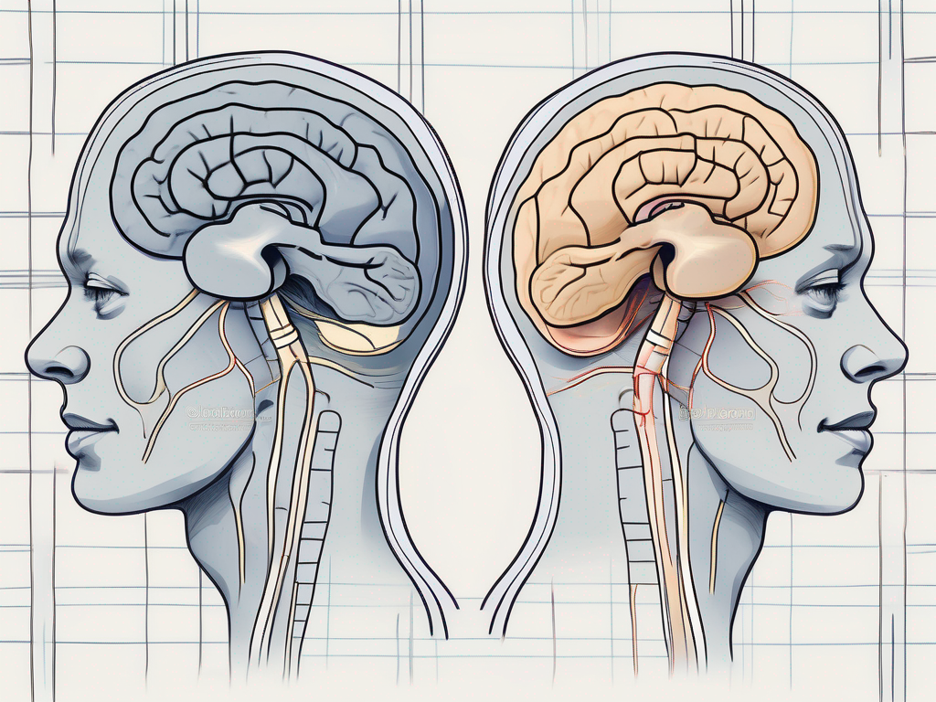
The vestibulocochlear nerve, also known as the eighth cranial nerve, plays a crucial role in our ability to hear and maintain balance. This remarkable nerve carries sensory information from the inner ear to the brain, where it is processed and interpreted. Understanding the intricacies of the vestibulocochlear nerve and its processing mechanisms is essential for comprehending the complexities of our auditory and vestibular systems. In this article, we will explore the anatomy, function, and connection of the vestibulocochlear nerve, as well as the disorders that can affect it.
Understanding the Vestibulocochlear Nerve
The vestibulocochlear nerve, also known as the eighth cranial nerve, is a crucial component of our auditory and vestibular systems. It plays a vital role in our ability to perceive and interpret sound, as well as maintain our balance and spatial orientation. Let’s delve deeper into the anatomy and function of this remarkable nerve.
Anatomy of the Vestibulocochlear Nerve
The vestibulocochlear nerve is composed of two distinct branches: the vestibular branch and the cochlear branch. These branches work in harmony to provide us with a comprehensive understanding of our auditory and vestibular world.
The vestibular branch carries information related to balance and spatial orientation. It receives signals from the vestibular apparatus, which consists of the utricle, saccule, and semicircular canals located within the inner ear. These structures are responsible for detecting changes in head position and movement, allowing us to maintain our balance and coordinate our movements effortlessly.
On the other hand, the cochlear branch is responsible for transmitting auditory signals. It receives input from the cochlea, a spiral-shaped structure within the inner ear. The cochlea contains thousands of tiny hair cells that convert sound waves into electrical signals. These electrical signals are then transmitted through the cochlear branch of the vestibulocochlear nerve to the brainstem, where they are further processed and analyzed.
The intricate anatomy of the vestibulocochlear nerve ensures that both our balance and hearing systems work seamlessly together, allowing us to navigate the world around us with ease.
Function of the Vestibulocochlear Nerve
The vestibulocochlear nerve serves a vital role in our ability to perceive and interpret sound. When sound waves enter the ear, they travel through the ear canal and reach the eardrum. The eardrum vibrates in response to the sound waves, which sets the chain of events in motion.
These vibrations are then transmitted through the middle ear, where three small bones called the ossicles (malleus, incus, and stapes) amplify the sound waves. The amplified vibrations reach the cochlea, where the hair cells convert them into electrical signals.
These electrical signals are then transmitted through the cochlear branch of the vestibulocochlear nerve to the brainstem. In the brainstem, the signals are processed and analyzed by various auditory centers, allowing us to recognize various sounds, ranging from the melodious tunes of music to the gentle whispers of nature.
In addition to its role in hearing, the vestibulocochlear nerve also plays a crucial role in maintaining our balance. The vestibular branch of the nerve receives signals from the vestibular apparatus, which detects changes in head position and movement. These signals are then transmitted to the brainstem, where they are integrated with visual and proprioceptive information to maintain our balance and coordinate our movements.
Overall, the vestibulocochlear nerve is a remarkable structure that allows us to experience the rich tapestry of sounds and maintain our equilibrium in the world around us. Its intricate anatomy and complex function highlight the intricate design of the human body and the wonders of our auditory and vestibular systems.
The Brain’s Role in Processing Vestibulocochlear Information
The Auditory Pathway: From Ear to Brain
Once the vestibulocochlear nerve carries auditory signals from the cochlea to the brainstem, they continue their journey along the auditory pathway. This intricate network of neural connections passes through several areas within the brain, each contributing to the processing and interpretation of sound. From the brainstem, the auditory signals ascend to the midbrain, thalamus, and finally reach their destination in the auditory cortex.
The midbrain, also known as the mesencephalon, is a vital part of the auditory pathway. It acts as a relay station, transmitting the auditory signals from the brainstem to the thalamus. Additionally, the midbrain plays a role in sound localization, helping us determine the direction from which a sound is coming.
As the auditory signals reach the thalamus, another crucial step in sound processing occurs. The thalamus acts as a gateway, filtering and prioritizing the auditory information before it is sent to the auditory cortex. This selective processing ensures that only relevant sounds are given attention, while irrelevant background noise is filtered out.
Finally, the auditory signals reach their destination in the auditory cortex, located within the temporal lobe of the brain. The auditory cortex is divided into different regions, each specializing in different aspects of sound processing. For example, the primary auditory cortex is responsible for basic sound analysis, such as distinguishing different frequencies. On the other hand, higher-order regions of the auditory cortex are involved in more complex tasks, such as recognizing speech patterns and assigning meaning to the auditory information received.
The Role of the Auditory Cortex
The auditory cortex, located within the temporal lobe of the brain, plays a crucial role in making sense of the sounds we perceive. It is responsible for distinguishing different frequencies, recognizing speech patterns, and assigning meaning to the auditory information received. Without the intricate network of neural connections and the processing capabilities of the auditory cortex, our experience of sound would be diminished.
Within the auditory cortex, there are specialized cells called “tonotopic maps” that are responsible for organizing and processing different frequencies. These maps allow us to distinguish between high-pitched sounds, like a bird’s chirping, and low-pitched sounds, like a rumbling thunder.
Furthermore, the auditory cortex is involved in auditory memory. It helps us recognize familiar sounds, such as the voice of a loved one or a favorite song. This memory aspect of the auditory cortex is essential for our ability to navigate the auditory world and make meaningful connections with the sounds we encounter.
In addition to its role in sound processing, the auditory cortex also interacts with other areas of the brain to integrate auditory information with other sensory modalities. For example, it collaborates with the visual cortex to help us locate the source of a sound by combining auditory cues with visual cues. This integration of sensory information enhances our ability to perceive and interact with the environment.
In conclusion, the brain’s processing of vestibulocochlear information involves a complex network of neural connections and specialized regions, such as the auditory cortex. Through this intricate system, we are able to perceive and make sense of the sounds around us, enriching our experience of the auditory world.
The Connection between the Vestibulocochlear Nerve and Balance
The Vestibular System Explained
In addition to its crucial role in hearing, the vestibulocochlear nerve also forms a vital connection with our sense of balance. The vestibular system, located within the inner ear, works in conjunction with the vestibulocochlear nerve to provide us with a stable and coordinated sense of equilibrium. It helps us maintain an upright posture, detects head movements, and contributes to our ability to walk, run, and perform everyday activities without losing our balance.
The vestibular system consists of two main components: the semicircular canals and the otolith organs. The semicircular canals are three fluid-filled structures that are responsible for detecting rotational movements of the head. Each canal is oriented in a different plane, allowing us to perceive movement in all directions. The otolith organs, on the other hand, detect linear acceleration and changes in head position relative to gravity. They contain tiny calcium carbonate crystals called otoliths, which move in response to changes in head position and stimulate hair cells that send signals to the brain.
When we move our heads, the fluid in the semicircular canals and the movement of the otoliths in the otolith organs cause the hair cells to bend. This bending generates electrical signals that are transmitted through the vestibulocochlear nerve to the brain. The brain then processes these signals and uses them to make adjustments to our muscle activity, allowing us to maintain our balance.
How Balance is Maintained
Balance is the result of a complex interplay between the vestibular system, visual input, and proprioception (the ability to perceive the position and movement of our body parts). When the vestibulocochlear nerve detects changes in head position or movement, it sends this information to the brain, which then modifies muscle activity to maintain balance. This intricate feedback loop ensures that our movements remain controlled and coordinated.
Visual input plays a crucial role in maintaining balance. Our eyes provide information about the position of our body in relation to the environment, allowing us to make adjustments to our posture and movements. For example, if we are walking on an uneven surface, our eyes can detect the changes in the terrain and send signals to the brain, which then adjusts our muscle activity to prevent us from losing our balance.
Proprioception, the ability to sense the position and movement of our body parts, also contributes to our sense of balance. Specialized receptors called proprioceptors are located in our muscles, tendons, and joints. These receptors provide information about the position of our limbs and the amount of tension in our muscles. This information is then integrated with the signals from the vestibular system and visual input to maintain balance.
When there is a disruption in any of these systems, such as damage to the vestibulocochlear nerve or a loss of visual input, it can result in balance problems. People with vestibular disorders may experience dizziness, vertigo, unsteadiness, and difficulty with coordination. Treatment for balance disorders often involves a combination of physical therapy, medication, and lifestyle modifications to improve overall balance and reduce symptoms.
Disorders Associated with the Vestibulocochlear Nerve
The vestibulocochlear nerve, also known as the eighth cranial nerve, plays a crucial role in our ability to hear and maintain balance. It is a remarkable structure that consists of two branches – the vestibular branch, responsible for balance and spatial orientation, and the cochlear branch, responsible for hearing. When this nerve is disrupted or damaged, it can lead to a variety of symptoms that can significantly impact an individual’s quality of life.
Symptoms of Vestibulocochlear Nerve Disorders
One of the most common symptoms associated with vestibulocochlear nerve disorders is hearing loss. This can range from mild to severe and may affect one or both ears. Individuals with hearing loss may struggle to understand conversations, hear certain frequencies, or experience a constant ringing sensation in their ears, known as tinnitus.
In addition to hearing loss, vestibulocochlear nerve disorders can also cause dizziness and vertigo. Dizziness refers to a sensation of lightheadedness or unsteadiness, while vertigo is characterized by a spinning or whirling sensation. These symptoms can be debilitating and may lead to difficulties with balance and coordination.
Imbalance is another common symptom of vestibulocochlear nerve disorders. Individuals may feel unsteady on their feet, have difficulty walking in a straight line, or experience frequent falls. This can greatly impact their day-to-day activities and overall quality of life.
If you experience any of these symptoms, it is important to seek medical attention to determine the underlying cause and explore appropriate treatment options. A healthcare professional specializing in audiology and vestibular disorders can conduct a comprehensive evaluation to diagnose the specific disorder and recommend the most suitable course of action.
Diagnosis and Treatment Options
Diagnosing vestibulocochlear nerve disorders typically involves a thorough evaluation by a healthcare professional. This evaluation may include a detailed medical history, physical examination, hearing tests, and imaging studies to pinpoint the precise location and nature of the problem.
Treatment options for vestibulocochlear nerve disorders vary depending on the specific disorder and its severity. In some cases, medication may be prescribed to manage symptoms such as dizziness or tinnitus. Vestibular rehabilitation, a form of physical therapy, can help improve balance and reduce symptoms of vertigo. Hearing aids may be recommended for individuals with hearing loss, while surgical interventions may be necessary for certain conditions.
It is crucial to consult with a qualified healthcare professional to discuss the most appropriate course of action for your individual needs. They can provide personalized guidance and support throughout your treatment journey, helping you regain control over your auditory and vestibular health.
In conclusion, the vestibulocochlear nerve is a remarkable structure that allows us to perceive sound and maintain balance. Its intricate anatomy and complex processing mechanisms ensure that we can appreciate the rich tapestry of sounds that surround us while moving through the world with grace and stability. By understanding the role of the vestibulocochlear nerve and recognizing the signs and symptoms of associated disorders, we can seek timely assistance from medical professionals and embark on a journey towards improved auditory and vestibular health.
