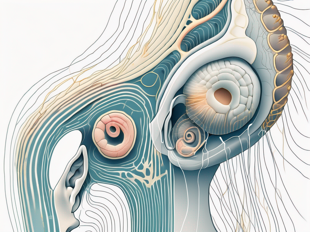
The complex process of hearing involves numerous structures and pathways within the ear. One important component in this process is the cochlea, which houses the receptors responsible for transmitting sounds to the vestibulocochlear nerve. In this article, we will explore the function of the cochlea, the role of the vestibulocochlear nerve, the types of receptors present in the cochlea, and the connection between the cochlea and the vestibulocochlear nerve. Additionally, we will discuss common disorders related to these structures and conclude with a recap of the intricate process of hearing.
Understanding the Function of the Cochlea
The cochlea is a spiral-shaped structure located in the inner ear. Its primary function is to convert sound vibrations into electrical signals that can be interpreted by the brain. To better comprehend the role of the cochlea, let’s delve into its anatomy.
Anatomy of the Cochlea
The cochlea consists of three fluid-filled chambers: the scala vestibuli, the scala media, and the scala tympani. These chambers are separated by a thin, flexible membrane known as the basilar membrane. Embedded within the basilar membrane are the organ of Corti and its hair cells, which serve as the main cochlear receptors.
The scala vestibuli, the uppermost chamber of the cochlea, is filled with perilymph, a fluid similar in composition to cerebrospinal fluid. This fluid helps transmit sound vibrations from the middle ear to the inner ear. Adjacent to the scala vestibuli is the scala media, also known as the cochlear duct. Filled with endolymph, a fluid with a high potassium concentration, the scala media plays a crucial role in the transduction of sound.
The scala tympani, the lowermost chamber of the cochlea, is also filled with perilymph. It extends from the apex of the cochlea to the round window, a flexible membrane that allows for the dissipation of pressure waves. The scala tympani is separated from the scala media by the basilar membrane, which acts as a partition between the two chambers.
The organ of Corti, located on the basilar membrane, contains specialized sensory cells called hair cells. These hair cells are responsible for converting mechanical vibrations into electrical signals. The hair cells are arranged in rows along the length of the cochlea, with the inner hair cells located closer to the modiolus, the central core of the cochlea, and the outer hair cells situated towards the periphery.
Role of the Cochlea in Hearing
When sound waves enter the ear, they travel through the ear canal and cause the eardrum to vibrate. These vibrations are then transmitted to the middle ear bones, the ossicles, which amplify and transmit the sound energy to the cochlea. Within the cochlea, the movement of fluid in the scala media displaces the basilar membrane, causing the hair cells to bend.
The bending of the hair cells triggers the release of neurotransmitters, specifically glutamate, which stimulate the auditory nerve fibers located in close proximity. These nerve fibers, known as the cochlear nerve, ultimately transmit the electrical signals generated by the hair cells to the brainstem, where they are further processed and interpreted as sound.
It is important to note that the cochlea plays a crucial role in the perception of different frequencies. The basilar membrane, due to its varying stiffness along its length, responds differently to different frequencies of sound. High-frequency sounds cause maximum displacement of the basilar membrane near the base of the cochlea, while low-frequency sounds cause maximum displacement near the apex.
Furthermore, the outer hair cells in the cochlea play a significant role in amplifying soft sounds and enhancing the overall sensitivity of the auditory system. These cells can change their length in response to electrical signals, thereby amplifying the mechanical vibrations and improving the detection of faint sounds.
In conclusion, the cochlea is a complex and remarkable structure that allows us to perceive and interpret the sounds around us. Its anatomy, including the scala vestibuli, scala media, and scala tympani, along with the organ of Corti and its hair cells, work together to convert sound vibrations into electrical signals that can be understood by the brain. Understanding the function of the cochlea is essential in comprehending the intricate process of hearing.
Introduction to Vestibulocochlear Nerve
The vestibulocochlear nerve, also known as the eighth cranial nerve, plays a crucial role in sound transmission. Let’s explore its structure and function in more detail.
Structure of the Vestibulocochlear Nerve
The vestibulocochlear nerve consists of two branches: the vestibular branch and the cochlear branch. While the vestibular branch is responsible for carrying information related to balance and spatial orientation, the cochlear branch specifically transmits auditory information from the cochlea to the brain.
The vestibular branch originates from the vestibular ganglion, which is located within the inner ear. It receives signals from the semicircular canals, utricle, and saccule, which are responsible for detecting changes in head position and movement. These signals are then transmitted through the vestibular branch to the brain, allowing us to maintain balance and coordination.
On the other hand, the cochlear branch originates from the spiral ganglion, which is also located within the inner ear. This branch carries electrical signals generated by the cochlea’s hair cells, which are responsible for converting sound vibrations into neural impulses. These impulses are then transmitted through the cochlear branch to the brain for further processing and interpretation.
Function of the Vestibulocochlear Nerve in Sound Transmission
As the electrical signals generated by the cochlea’s hair cells reach the auditory nerve fibers, they are transmitted via the cochlear branch of the vestibulocochlear nerve. These signals travel along the nerve fibers to the brainstem, where they are further processed and relayed to the auditory centers of the brain for interpretation.
Within the brainstem, the auditory nerve fibers synapse with neurons in the cochlear nuclei. From there, the information is relayed to various auditory pathways, including the superior olivary complex, inferior colliculus, and medial geniculate nucleus. These structures play a crucial role in processing and integrating auditory information, allowing us to perceive and understand sounds.
It is important to note that any disruption or damage to the vestibulocochlear nerve can lead to hearing loss and other auditory impairments. There are various factors that can contribute to nerve damage, including exposure to loud noises, infections, tumors, and certain medical conditions. If you experience any concerning changes in your hearing, such as difficulty understanding speech or ringing in the ears, it is recommended to consult with a healthcare professional, such as an audiologist or an otolaryngologist, for accurate diagnosis and appropriate management strategies.
Receptors in the Cochlea
The cochlea, a spiral-shaped structure located in the inner ear, contains specific receptors known as hair cells, which are essential for sound transmission. Let’s explore the types of receptors present in the cochlea and how they function.
Types of Receptors in the Cochlea
There are two main types of hair cells in the cochlea: inner hair cells (IHCs) and outer hair cells (OHCs). While IHCs are primarily responsible for converting sound vibrations into electrical signals, OHCs play a crucial role in amplifying and fine-tuning these signals, enhancing the ear’s sensitivity to different frequencies.
Inner hair cells, numbering around 3,500 in humans, are arranged in a single row along the length of the cochlea. They are the primary sensory receptors and are responsible for transmitting the majority of auditory information to the brain. These cells have specialized structures called stereocilia, which are tiny hair-like projections that extend from their tops.
Outer hair cells, on the other hand, are more numerous, with approximately 12,000 in humans. They are arranged in three rows and are involved in the active process of amplifying sound signals. These cells possess a unique structure called the prestin protein, which allows them to change their shape rapidly in response to electrical signals. This shape change, in turn, enhances the movement of the basilar membrane, leading to increased sensitivity to soft sounds and improved frequency discrimination.
How Cochlear Receptors Transmit Sound
When sound waves enter the ear, they cause the basilar membrane, a flexible structure that runs along the length of the cochlea, to vibrate. This vibration is crucial for the functioning of the hair cells. As the basilar membrane moves, the hair cells, anchored at their bases, bend in response to this movement.
The bending of the hair cells leads to the opening of ion channels, specialized protein channels located on the stereocilia. These channels allow ions, such as potassium and calcium, to enter the cells, generating electrical signals. The movement of these ions triggers a series of biochemical events within the hair cells, ultimately resulting in the release of neurotransmitters.
These neurotransmitters, such as glutamate, are released from the hair cells and bind to receptors on the auditory nerve fibers, which are connected to the hair cells. This binding initiates a cascade of electrical impulses that travel along the auditory nerve fibers towards the brain.
The intricate interaction between the hair cells, the basilar membrane, and the fluid-filled chambers of the cochlea ensures the precise and efficient transmission of sound information. This remarkable process allows us to perceive and interpret the rich tapestry of sounds that surround us in our everyday lives.
The Connection between Cochlea and Vestibulocochlear Nerve
Now that we have explored the individual functions of the cochlea and the vestibulocochlear nerve, let’s delve deeper into their intricate connection and the fascinating process of sound transmission.
The pathway of sound from the cochlea to the vestibulocochlear nerve is a complex and remarkable journey. Once the electrical signals are generated by the hair cells in the cochlea, they embark on a mission to reach the brainstem, where they will be further processed and relayed to the auditory centers of the brain for interpretation.
The cochlear branch of the vestibulocochlear nerve serves as the messenger, carrying these signals from the cochlea to the brainstem. This branch is responsible for ensuring that the information encoded in the electrical signals is accurately transmitted and received by the brain.
As the signals travel along the cochlear branch, they encounter numerous checkpoints and relay stations within the brainstem. These checkpoints play a crucial role in refining and enhancing the signals, ensuring that they are ready for interpretation in the auditory centers of the brain.
Once the signals successfully navigate through the brainstem, they finally arrive at the auditory centers of the brain, where the magic happens. These centers, located in the temporal lobes, are responsible for transforming the electrical signals into meaningful sounds that we can perceive and understand.
It is truly remarkable how the cochlea and the vestibulocochlear nerve work together seamlessly to enable us to hear and interpret the world around us. The presence of functional cochlear receptors, specifically the inner and outer hair cells, is crucial for the accurate transmission of sound signals to the vestibulocochlear nerve.
The inner hair cells are responsible for converting sound vibrations into electrical signals, while the outer hair cells play a vital role in amplifying and fine-tuning these signals. Any abnormalities or damage to these receptors can lead to various hearing disorders and impairments, affecting our ability to fully experience the richness of sound.
If you suspect any issues with your hearing, it is advisable to seek professional evaluation to identify the underlying cause and determine appropriate treatment options. The intricate connection between the cochlea and the vestibulocochlear nerve reminds us of the delicate nature of our auditory system and the importance of taking care of our hearing health.
Disorders Related to Cochlea and Vestibulocochlear Nerve
While the cochlea and the vestibulocochlear nerve are intricate structures involved in the hearing process, they can be susceptible to various disorders. Let’s discuss some common disorders associated with these structures.
The cochlea, a spiral-shaped structure located in the inner ear, is responsible for converting sound vibrations into electrical signals that can be interpreted by the brain. The vestibulocochlear nerve, also known as the eighth cranial nerve, carries these electrical signals from the cochlea to the brain, allowing us to perceive and interpret sounds.
Common Disorders and Their Symptoms
Some examples of disorders related to the cochlea and the vestibulocochlear nerve include sensorineural hearing loss, tinnitus, and Meniere’s disease.
Sensorineural hearing loss is characterized by a decreased ability to perceive sounds, often caused by damage to the hair cells or the auditory nerve fibers. This type of hearing loss can be caused by various factors, including aging, exposure to loud noises, certain medications, and genetic predisposition. Individuals with sensorineural hearing loss may experience difficulty understanding speech, trouble hearing in noisy environments, and a sense of muffled or distorted sound.
Tinnitus refers to the perception of ringing or buzzing sounds in the absence of external stimuli. It can be a symptom of various underlying conditions, including damage to the cochlea, exposure to loud noises, ear infections, or certain medications. Tinnitus can range from a mild annoyance to a debilitating condition that affects sleep, concentration, and overall quality of life.
Meniere’s disease is a disorder that affects the inner ear, leading to symptoms such as vertigo, hearing loss, and tinnitus. It is believed to be caused by an abnormal buildup of fluid in the inner ear, which disrupts the normal balance and hearing mechanisms. Individuals with Meniere’s disease may experience sudden episodes of severe vertigo, accompanied by fluctuating hearing loss and a feeling of fullness or pressure in the affected ear.
If you experience any symptoms of these disorders, it is crucial to consult with a healthcare professional, preferably an audiologist or an otolaryngologist, for accurate diagnosis and appropriate management strategies.
Treatment and Management of These Disorders
The treatment and management of disorders related to the cochlea and the vestibulocochlear nerve vary depending on the specific condition and its underlying cause.
In cases of sensorineural hearing loss, hearing aids or cochlear implants may be recommended to improve hearing. These devices work by amplifying sound or directly stimulating the auditory nerve, bypassing the damaged hair cells in the cochlea. Additionally, assistive listening devices and communication strategies can help individuals with hearing loss navigate daily communication challenges.
Tinnitus management approaches can include sound therapy, which aims to mask or distract from the perceived ringing or buzzing sounds. Counseling and cognitive behavioral therapy techniques may also be beneficial in helping individuals cope with the emotional and psychological impact of tinnitus.
For individuals with Meniere’s disease, treatment options may include medications to control symptoms such as vertigo and nausea. Dietary changes, such as reducing salt intake, and lifestyle modifications, such as stress management and regular exercise, can also play a crucial role in managing the condition and reducing the frequency and severity of episodes.
It is essential to emphasize that seeking professional medical advice is paramount for an accurate diagnosis and personalized treatment plan. Self-diagnosis and self-medication can potentially exacerbate the condition or delay appropriate intervention. A healthcare professional will conduct a thorough evaluation, which may include a comprehensive audiological assessment, to determine the underlying cause of the disorder and develop an individualized treatment approach.
Conclusion: The Intricate Process of Hearing
In conclusion, the receptors found in the cochlea play a vital role in transmitting sounds to the vestibulocochlear nerve, enabling us to perceive and interpret the world of sound. The cochlear hair cells, along with the vestibulocochlear nerve, form an intricate system that ensures the accurate transmission of auditory information to the brain. Understanding the anatomy, function, and disorders related to these structures enhances our appreciation of the complexity of the hearing process.
Recap of Cochlear Receptors and Vestibulocochlear Nerve
The cochlear receptors, including inner and outer hair cells, are responsible for converting sound vibrations into electrical signals. These signals are then transmitted to the brain via the vestibulocochlear nerve, specifically through its cochlear branch. The accurate transmission of sound information relies on the coordinated functioning of these structures.
Future Research Directions in Auditory Neuroscience
Advancements in auditory neuroscience continue to shed light on the intricate mechanisms underlying the hearing process. Ongoing research aims to further our understanding of cochlear receptors, the vestibulocochlear nerve, and the interplay between these structures. This ever-evolving field holds the promise of developing innovative diagnostic techniques and targeted therapies for hearing disorders, ultimately improving the quality of life for individuals affected by auditory impairments.
