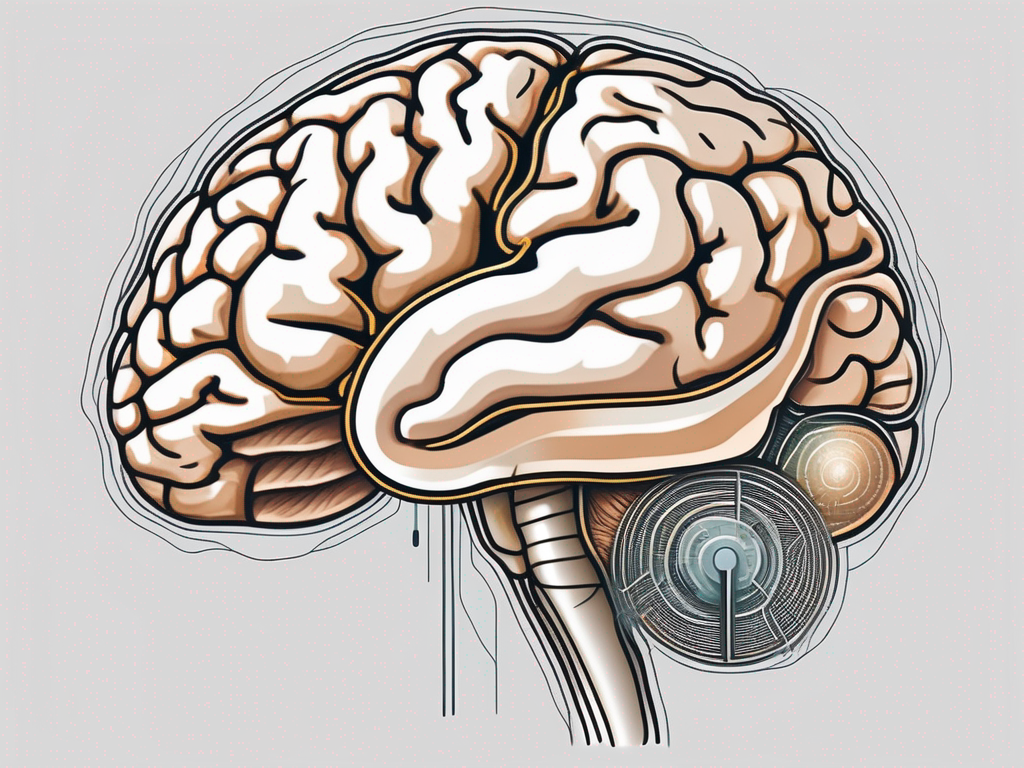
The vestibulocochlear nerve, also known as the eighth cranial nerve, plays a crucial role in our ability to hear and maintain balance. It is responsible for transmitting sensory information from the inner ear to the brain, where it is processed and interpreted. Understanding the intricate connection between the vestibulocochlear nerve and the brain is essential to comprehend how we perceive sound and maintain our sense of equilibrium.
Understanding the Vestibulocochlear Nerve
The vestibulocochlear nerve is a paired nerve that consists of two distinct branches: the vestibular branch and the cochlear branch. The vestibular branch is associated with the sense of balance and spatial orientation, while the cochlear branch is dedicated to hearing. Together, they relay vital information to the brain, allowing us to perceive sound and maintain our stability in the surrounding environment.
Anatomy of the Vestibulocochlear Nerve
The vestibulocochlear nerve originates from the inner ear, which comprises the cochlea and vestibular apparatus. The cochlea is responsible for converting sound vibrations into electrical signals, while the vestibular apparatus senses changes in head position and movement. These specialized structures send signals through the respective branches of the vestibulocochlear nerve, ultimately reaching the brain for further processing.
The cochlea, often referred to as the “snail-shaped” structure, is a complex organ that plays a crucial role in our ability to hear. It is lined with thousands of tiny hair cells that are responsible for converting sound waves into electrical signals. These hair cells are incredibly sensitive and can detect even the slightest variations in sound frequency and intensity.
Adjacent to the cochlea lies the vestibular apparatus, which consists of three semicircular canals and two otolith organs. The semicircular canals are responsible for detecting rotational movements of the head, while the otolith organs sense linear acceleration and changes in head position. These structures work in harmony to provide us with a sense of balance and spatial orientation.
Function of the Vestibulocochlear Nerve
The vestibulocochlear nerve serves as the primary pathway for auditory and vestibular information to travel from the ear to the brain. The vestibular branch transmits signals related to balance, providing us with a sense of spatial orientation and helping us maintain stability. This branch is responsible for detecting head movements, whether it be tilting, rotating, or accelerating, and relaying this information to the brain.
When we walk, run, or engage in any physical activity, the vestibular branch of the vestibulocochlear nerve is constantly at work, ensuring that our body maintains its equilibrium. It allows us to adjust our posture and make necessary adjustments to prevent falls or accidents.
The cochlear branch of the vestibulocochlear nerve, on the other hand, carries electrical signals that represent sound waves. These signals are generated by the hair cells in the cochlea and are then transmitted to the brain for interpretation. The brain decodes these signals, allowing us to perceive and interpret different sounds, whether it be the chirping of birds, the sound of music, or the voice of a loved one.
Without the vestibulocochlear nerve, our ability to hear and maintain balance would be severely compromised. It is through this remarkable nerve that we are able to navigate the world around us, enjoying the symphony of sounds and staying steady on our feet.
The Brain and its Various Parts
The brain, a complex and intricate organ, plays a crucial role in processing the information received from the vestibulocochlear nerve. It consists of multiple interconnected regions, each responsible for different aspects of perception, cognition, and behavior.
The brain, being the command center of the nervous system, is a fascinating organ with a multitude of functions. Let’s delve deeper into the structure and functions of its various parts.
Overview of the Brain’s Structure
The brain can be broadly categorized into three main regions: the forebrain, midbrain, and hindbrain. Each of these regions has its own unique characteristics and functions.
The forebrain, also known as the cerebrum, is the largest part of the brain and is responsible for complex cognitive functions. It is divided into two hemispheres, the left and the right, which are connected by a bundle of nerve fibers called the corpus callosum. The cerebrum is further divided into lobes, including the frontal lobe, parietal lobe, temporal lobe, and occipital lobe. These lobes play a crucial role in processes such as decision-making, sensory perception, language comprehension, and visual processing.
The midbrain acts as a relay station, transmitting information between the forebrain and the hindbrain. It plays a vital role in coordinating movements, regulating sleep-wake cycles, and controlling involuntary reflexes. The midbrain also contains structures such as the substantia nigra, which produces dopamine, a neurotransmitter involved in movement and reward.
The hindbrain, composed of the pons, medulla oblongata, and the cerebellum, controls essential bodily functions such as heart rate, breathing, and coordination. The pons serves as a bridge between the cerebrum and the cerebellum, while the medulla oblongata regulates vital functions like breathing, blood pressure, and heart rate. The cerebellum, often referred to as the “little brain,” is responsible for fine motor control, balance, and coordination.
Role of Different Brain Parts in Processing Information
Several specific regions within the brain are involved in processing the sensory information transmitted by the vestibulocochlear nerve. Let’s explore some of these regions and their functions.
The temporal lobe, located on the sides of the brain, plays a vital role in auditory processing. It receives sound signals from the cochlear branch and further analyzes and interprets them. The temporal lobe is not only responsible for basic sound perception but also for more complex auditory functions, such as recognizing and understanding speech, music, and other sounds. It is also involved in memory formation and retrieval, particularly for auditory memories.
Within the temporal lobe lies the auditory cortex, a region dedicated to processing auditory information. The auditory cortex receives input from the cochlea and performs intricate computations to assign meaning to the sounds we hear. It helps us distinguish between different pitches, tones, and frequencies, allowing us to recognize and interpret a wide range of sounds in our environment.
Furthermore, the auditory cortex is connected to other areas of the brain, such as the language centers in the left hemisphere. This connection enables us to understand spoken language and engage in meaningful conversations. It also allows us to appreciate the beauty of music and enjoy the emotional impact it can have on our lives.
In conclusion, the brain is an incredibly complex organ, consisting of various interconnected regions that work together to process information and regulate our thoughts, emotions, and behaviors. Understanding the structure and functions of these brain parts helps us appreciate the intricate workings of the human mind.
The Pathway of the Vestibulocochlear Nerve to the Brain
The journey of the vestibulocochlear nerve from the inner ear to the brain involves a complex pathway, facilitating the transmission of sensory information in real-time.
The vestibulocochlear nerve, also known as the eighth cranial nerve, is responsible for carrying auditory and vestibular information from the inner ear to the brain. This nerve plays a crucial role in our ability to hear and maintain balance.
The Journey from the Inner Ear to the Brain
After sound vibrations are detected by the cochlea, they are converted into electrical signals. From there, the cochlear branch of the vestibulocochlear nerve relays these signals to the brainstem, an integral part of the midbrain. The brainstem acts as a crucial junction, connecting the vestibulocochlear nerve to various regions of the brain responsible for processing auditory and balance-related information.
As the electrical signals travel along the cochlear branch, they pass through a series of specialized cells called spiral ganglion cells. These cells are located within the cochlea and serve as the first relay station for auditory information. The spiral ganglion cells convert the electrical signals into neural impulses, which are then transmitted to the brainstem.
Once the neural impulses reach the brainstem, they undergo further processing and integration. This processing involves the coordination of auditory and vestibular information to ensure proper balance and spatial orientation. The brainstem acts as a gateway, directing the neural impulses to the appropriate brain regions for further analysis and interpretation.
The Role of the Brainstem in Vestibulocochlear Nerve Function
The brainstem acts as a relay center, receiving and processing sensory information from the vestibulocochlear nerve before further relaying it to the appropriate brain regions. This includes relaying auditory information to the temporal lobe, where the brain processes sound signals and assigns meaning to them. The brainstem’s involvement in vestibulocochlear nerve function highlights its vital role in maintaining balance and processing auditory stimuli.
In addition to its role in auditory processing, the brainstem also plays a crucial role in regulating balance and spatial orientation. It receives vestibular information from the vestibular branch of the vestibulocochlear nerve, which is responsible for detecting changes in head position and movement. This information is essential for maintaining equilibrium and coordinating body movements.
Furthermore, the brainstem is involved in the reflexive responses that occur in response to auditory and vestibular stimuli. For example, when we hear a sudden loud noise, the brainstem initiates a startle reflex, causing our body to react quickly. Similarly, when we experience a sudden change in head position, the brainstem triggers the vestibulo-ocular reflex, which helps to stabilize our gaze and maintain visual focus.
Overall, the pathway of the vestibulocochlear nerve from the inner ear to the brain is a complex and intricate process. It involves multiple stages of signal transmission, processing, and integration, all of which are essential for our ability to hear, maintain balance, and navigate the world around us.
The Brain Part Receiving Information from the Vestibulocochlear Nerve
The temporal lobe, located within the cerebrum, is the primary brain region responsible for processing sound information received from the cochlear branch of the vestibulocochlear nerve.
The temporal lobe, a fascinating and intricate part of the brain, plays a crucial role in our ability to hear and interpret sound. Nestled within the cerebrum, this region is specifically dedicated to processing the electrical signals that represent sound waves. These signals are received from the cochlear branch of the vestibulocochlear nerve, a vital pathway for auditory information.
The Role of the Temporal Lobe in Hearing
The temporal lobe houses the auditory cortex, which is dedicated to processing sound information. It receives electrical signals representing sound waves from the vestibulocochlear nerve’s cochlear branch and decodes them, allowing us to discern different frequencies, volumes, and qualities of sound.
Within the temporal lobe, an intricate network of neurons and specialized areas work harmoniously to ensure the accurate interpretation and understanding of auditory stimuli. This remarkable process involves the coordination of various brain regions, each contributing to our ability to perceive and make sense of the sounds around us.
How the Auditory Cortex Processes Sound Information
The auditory cortex within the temporal lobe is responsible for various aspects of auditory processing, including sound localization, pitch recognition, and speech comprehension. Neurons in the auditory cortex analyze the received sound signals and extract relevant information such as the direction sound is coming from or the meaning behind spoken words.
As the auditory cortex processes sound information, it engages in a complex dance of neural activity. Different regions within the auditory cortex specialize in specific aspects of sound processing, allowing for a highly nuanced understanding of auditory stimuli. This intricate web of neural connections enables us to not only hear sounds but also to interpret their meaning and significance.
Sound localization, for example, is a remarkable ability facilitated by the auditory cortex. By analyzing subtle differences in the timing and intensity of sound reaching each ear, the brain can accurately determine the direction from which a sound originates. This remarkable feat is made possible by the precise coordination of neurons within the auditory cortex.
Pitch recognition, another essential function of the auditory cortex, allows us to distinguish between high and low-frequency sounds. This ability is crucial for understanding music, recognizing voices, and perceiving the emotional nuances conveyed through changes in pitch.
Speech comprehension, perhaps one of the most remarkable feats of the auditory cortex, involves the decoding of spoken words and the extraction of their meaning. The auditory cortex analyzes the intricate patterns of sound produced during speech, allowing us to understand and interact with the spoken language.
Overall, the temporal lobe and its auditory cortex are remarkable structures that contribute to our ability to hear and make sense of the auditory world. Through their intricate neural networks and specialized functions, they allow us to perceive, interpret, and appreciate the rich tapestry of sounds that surround us.
Disorders Related to the Vestibulocochlear Nerve
Although the vestibulocochlear nerve plays a critical role in hearing and maintaining balance, it is susceptible to disorders that can impair its function.
The vestibulocochlear nerve, also known as the eighth cranial nerve, is responsible for transmitting sensory information related to hearing and balance from the inner ear to the brain. This intricate network of nerve fibers allows us to perceive sound and maintain our sense of equilibrium. However, like any other part of the body, the vestibulocochlear nerve can be affected by various disorders that disrupt its normal functioning.
Symptoms of Vestibulocochlear Nerve Damage
Damage to the vestibulocochlear nerve can result in various symptoms. One of the most common symptoms is hearing loss, which can range from mild to severe. Individuals with vestibulocochlear nerve damage may experience difficulty hearing sounds or understanding speech. In addition to hearing loss, tinnitus, a condition characterized by the perception of ringing or buzzing sounds in the ears, can also occur.
Another symptom associated with vestibulocochlear nerve damage is dizziness. Individuals may feel a spinning sensation, known as vertigo, which can be accompanied by nausea and vomiting. Difficulties with balance and coordination are also common, making it challenging to walk or perform daily activities without assistance.
If you experience any of these symptoms, it is essential to consult with a healthcare professional for a comprehensive evaluation and appropriate management. Early detection and intervention can significantly improve outcomes and prevent further deterioration of the vestibulocochlear nerve function.
Treatment and Management of Vestibulocochlear Nerve Disorders
The specific treatment and management options for vestibulocochlear nerve disorders depend on the underlying cause and severity of the condition. In some cases, medical interventions may be necessary, including medications or surgical procedures. For example, if the damage is caused by an infection, antibiotics or antiviral drugs may be prescribed to treat the underlying infection and reduce inflammation.
However, not all vestibulocochlear nerve disorders require invasive treatments. In many cases, non-invasive approaches can be effective in managing the symptoms and improving quality of life. Hearing aids are commonly used to amplify sounds and compensate for hearing loss. These devices can be customized to fit individual needs and provide significant benefits in enhancing auditory perception.
Balance exercises, also known as vestibular rehabilitation, can help individuals with vestibulocochlear nerve disorders improve their balance and coordination. These exercises are designed to strengthen the vestibular system and promote adaptation to changes in body position and movement. They are often performed under the guidance of a physical therapist or an audiologist specialized in vestibular rehabilitation.
Lifestyle modifications can also play a crucial role in managing vestibulocochlear nerve disorders. Avoiding triggers that worsen symptoms, such as excessive noise or sudden head movements, can help minimize discomfort and improve overall well-being. Stress management techniques, such as relaxation exercises or meditation, may also be beneficial in reducing the impact of vestibulocochlear nerve-related symptoms.
In conclusion, the vestibulocochlear nerve plays a vital role in transmitting sensory information related to hearing and balance from the inner ear to the brain. Understanding the intricate connection between the vestibulocochlear nerve and the brain allows us to comprehend how we perceive sound and maintain our sense of equilibrium. However, the complexity of this system also makes it susceptible to disruptions and disorders that can affect our hearing and balance.
If you experience any symptoms related to the vestibulocochlear nerve, it is crucial to seek medical advice and guidance from healthcare professionals specializing in audiology or neurology. With proper evaluation and management, individuals with vestibulocochlear nerve disorders can regain or enhance their auditory and balance functions, improving their overall quality of life.
